Prediction Workflow
Follow the steps below to upload a urine microscopy image, run both models, and interpret the resulting UTI likelihood. The dialog is always available so you can test multiple samples without leaving this page.
Step-by-Step Guide
1. Prepare Sample
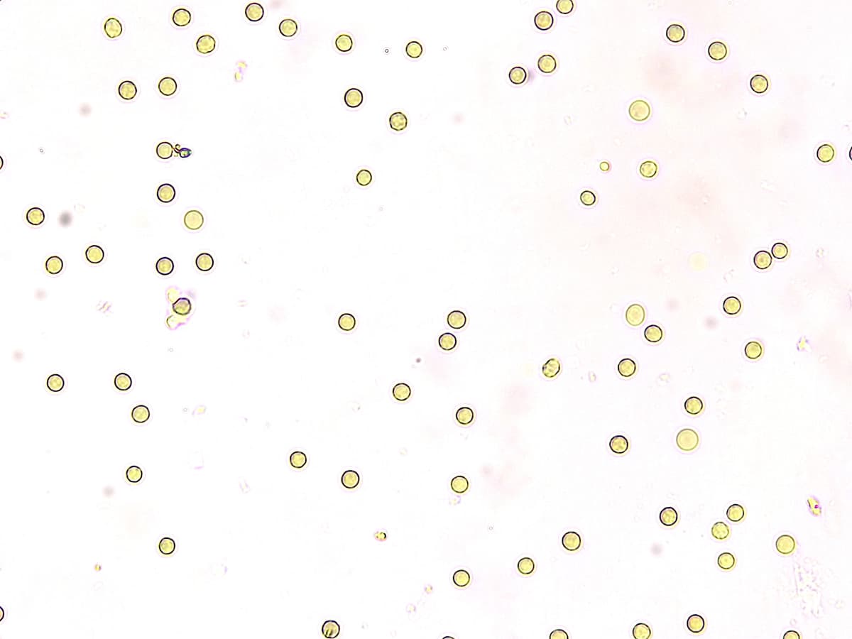
Capture or select a microscopic urine image that clearly shows cellular structures. Supported formats include PNG and JPEG.
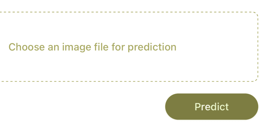
2. Upload Image
Use the upload dialog to select your file. The preview confirms the image was attached successfully before analysis.
3. Run Prediction
Tap Predict to run both Non-UTI and UTI classifiers sequentially. You can cancel mid-process if you selected the wrong file.
4. Review Results
Final probabilities and class labels appear in the results dialog. Re-run with another image anytime.
Example Screenshot